

Author: Kasem Jamil Al-Saloumi

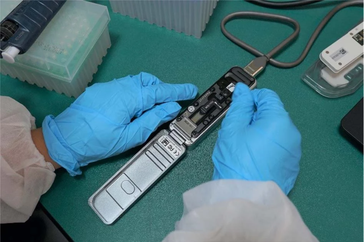
A portable MinION DNA sequencer being prepared for sequencing. Illustrative photograph; not from the author’s study. Credit: Mongan A.E., via Wikimedia Commons; CC BY 4.0 (https://creativecommons.org/licenses/by/4.0/). Source: https://commons.wikimedia.org/wiki/File:MinION_portable_sequencer.png. Image unmodified.
The first time a DNA sequencer gave me the name of a bacterium from a baby’s blood sample, I felt something close to pride. Then I felt uneasy. As it turned out, that unease was the more useful feeling.
In my postgraduate research, I worked with blood samples from premature infants who had been investigated for sepsis — a potentially life-threatening whole-body response to infection. Some had negative blood cultures, meaning that no organism grew under the laboratory’s incubation conditions. A negative culture does not necessarily rule out infection, which is why molecular tests are studied as complementary tools. These include polymerase chain reaction (PCR) (a technique that amplifies a specific DNA sequence) and sequencing methods that directly look for microbial genetic material rather than waiting for a bacterial organism to grow [1].
Our question was simple to ask but difficult to answer: could a portable DNA sequencer detect bacterial DNA that conventional blood culture had not detected?
The machine we used is small enough to sit next to a laptop. Once prepared DNA is loaded into it, the sequencer begins producing reads within hours: long stretches of DNA represented by strings of letters A, T, C and G. The software then compares each read with sequences from known organisms in a reference database and reports possible matches.
A genus is a broad group of closely related bacteria, while a species is a more specific identity within a genus. Depending on the read quality, the sequence being examined and the reference database, the software may identify a bacterium at the genus level and sometimes at the species level.
It looks like an answer. It is very tempting to treat it as one.
Here is the problem. Blood samples can contain very little microbial material. Scientists describe these as low-biomass samples. When the signal we are looking for is faint, traces of DNA from other sources matter much more. An extraction kit — a collection of materials and chemicals used to break open cells and extract and purify DNA — can itself contain bacterial DNA. Unwanted DNA can also be introduced through commercial reagents, water, handling, or the surrounding laboratory environment [2,3].
If a sample contains abundant bacterial DNA, a small amount of contaminating DNA may account for only a tiny fraction of the reads detected. In contrast, when genuine bacterial DNA is scarce, the same background contamination can make up a much larger fraction of what the sequencer detects. A weak contaminating signal can then look disproportionately important [2,3].
This is a recognised problem in low-biomass sequencing. Bacteria such as Ralstonia, Pseudomonas and Burkholderia have been reported as reagent-associated contaminants in microbiome studies [2,3]. This does not mean that every detection of these bacteria is contamination. Rather, it means that a plausible-looking result can sometimes reflect DNA introduced during laboratory processing. That possibility needs to be tested before drawing conclusions.
So what should you do when your sequencer reports, with an impressively precise match percentage, that a baby’s blood contains a particular bacterium?
One important step is to run negative controls, often called blanks.
A blank is a sample that goes through the same laboratory procedures as the real samples but contains no patient material. Blanks help researchers detect patterns that may indicate DNA introduced during extraction, amplification or other processing steps. They cannot, however, establish the origin of every sequence.
For instance, if the same bacterial sequence appears in both the patient’s sample and the corresponding blank, especially at a similar or higher level in the blank, contamination becomes a plausible alternative explanation [2,3].
However, blanks do not solve everything. Contamination can vary between samples, while a genuine infection may produce so little bacterial DNA that its signal is difficult to distinguish from background DNA. A possible solution for this problem could be using a software package, such as Decontam. Decontam uses statistical patterns to flag sequences that are more likely to be contaminants. For example, a sequence may appear more often in blanks or become more prominent as the total amount of sample DNA falls. These approaches have assumptions and limitations. They help researchers interpret the evidence, but cannot determine whether a detected organism actually caused a patient’s illness [4]. In borderline cases, there may still be insufficient evidence for a confident answer.
Even when contamination is unlikely, detecting bacterial DNA does not, by itself, prove that the organism caused a particular patient’s illness. This remains true even when the species is known to be pathogenic — capable of causing disease [5]. Reaching that conclusion depends on clinical context, including symptoms, timing, culture results, inflammatory markers, other laboratory findings (e.g. white blood cell counts) and prior antibiotic exposure, together with controls that test alternative explanations, especially contamination.
The reference database introduces another complication. The software can compare a DNA read only with sequences represented in the reference database used. Missing organisms, incorrect labels and highly similar sequences can affect the identification or limit it to a broader group, such as a genus [6,7].
This matters for the method used in my project: 16S sequencing examines a bacterial marker gene — a short stretch of DNA used to help identify bacteria — rather than every gene in the organism. Some closely related bacteria can have very similar versions of this marker. Even with a suitable database, the sequence may therefore contain too little information to distinguish confidently between species [8].
None of this means DNA sequencing is unreliable. It means we need to be clear about what a result can tell us.
A DNA read can resemble the sequence of a known organism without establishing whether it came from living or dead cells. Researchers sometimes use propidium monoazide, a dye that binds preferentially to DNA from cells with damaged membranes and reduces its amplification, to limit signals from dead bacteria [9]. A study of saliva found that this treatment changed the bacterial profiles obtained by 16S sequencing [10]. However, evidence from saliva does not validate the method for testing neonatal blood samples. Its performance still needs evaluation for the organisms and samples being studied.
The most valuable thing my postgraduate sequencing work taught me was to change the question I asked of each result. I stopped asking only, “What did we find?” and started asking, “What other explanation could produce this result if the target bacterium were absent?” If a result remains convincing after contaminating DNA, background DNA and other alternative explanations have been considered, it earns greater confidence. If it does not, the evidence may not be strong enough to support the original conclusion.
This lesson extends beyond the laboratory. DNA evidence carries considerable authority in courtrooms, hospitals and news headlines, partly because it appears so precise. A string of letters can feel more objective than a doctor’s judgment or a witness’s memory.
Precision and truth are different things, though. A measurement can be extremely precise and still be interpreted incorrectly, or reflect something other than what we intended to measure.
Scientists working with low-biomass samples can use several safeguards. Negative controls should accompany the samples, and interpretation criteria should be established and tested for the assay. A minimum read count or proportion of reads alone cannot distinguish contamination from a genuine signal because a contaminant can exceed that threshold. Researchers should also compare the organism’s pattern across samples and blanks, and consider independent evidence such as culture results, targeted PCR (a test designed to detect a specific organism or genetic target) and the patient’s clinical findings [2–4]. Recording the reference database and its release or version used is equally important. Databases are updated as sequences are added or corrected, and these changes can affect which organism a read is assigned to [6,7]. Finally, scientists should be communicate clearly what their results cannot establish, including whether a signal reflects contamination or DNA from non-viable cells [5,9].
Therefore, careful reporting matters. These limitations should be carried into public explanations rather than leaving them buried in a paper’s methods and results. A news story or social-media summary should distinguish detection from diagnosis, mention the relevant controls and explain important alternative interpretations. This lets scientists, clinicians and readers judge how much confidence to place in a finding.
If there is one thing I hope readers take away from this, it is that an impressive result deserves careful scrutiny before it is celebrated. That is not pessimism; it is how confidence is built. Careful scrutiny does not weaken a convincing result. It helps show which signals genuinely deserve our trust.
Article written by Kasem Jamil Al-Saloumi, who holds an MSc in Biotechnology (DNA Technology). His postgraduate research, conducted with the Biomedical Research Center at Qatar University, used Oxford Nanopore sequencing on blood samples from premature infants. He has taught biology for more than thirteen years and lives in Doha.
Article written by Priscilla Wong, a recent BSc Biological Sciences (Immunology) (Hons) graduate from the University of Edinburgh and the Head Online News Editor for EUSci.
References:
- Dierikx TH, Visser DH, de Meij T, et al. Molecular assays for the diagnosis of sepsis in neonates: a diagnostic test accuracy review. Cochrane Database of Systematic Reviews. 2025;3:CD011926. doi:10.1002/14651858.CD011926.pub3
- Salter SJ, Cox MJ, Turek EM, et al. Reagent and laboratory contamination can critically impact sequence-based microbiome analyses. BMC Biology. 2014;12:87. doi:10.1186/s12915-014-0087-z
- Eisenhofer R, Minich JJ, Marotz C, et al. Contamination in low microbial biomass microbiome studies: issues and recommendations. Trends in Microbiology. 2019;27(2):105–117. doi:10.1016/j.tim.2018.11.003
- Davis NM, Proctor DM, Holmes SP, Relman DA, Callahan BJ. Simple statistical identification and removal of contaminant sequences in marker-gene and metagenomics data. Microbiome. 2018;6:226. doi:10.1186/s40168-018-0605-2
- Chiu CY, Miller SA. Clinical metagenomics. Nature Reviews Genetics. 2019;20:341–355. doi:10.1038/s41576-019-0113-7
- Marcelino VR, Holmes EC, Sorrell TC. The use of taxon-specific reference databases compromises metagenomic classification. BMC Genomics. 2020;21:184. doi:10.1186/s12864-020-6592-2
- Chorlton SD. Ten common issues with reference sequence databases and how to mitigate them. Frontiers in Bioinformatics. 2024;4:1278228. doi:10.3389/fbinf.2024.1278228
- Johnson JS, Spakowicz DJ, Hong BY, et al. Evaluation of 16S rRNA gene sequencing for species and strain-level microbiome analysis. Nature Communications. 2019;10:5029. doi:10.1038/s41467-019-13036-1
- Kobayashi H, Oethinger M, Tuohy MJ, Hall GS, Bauer TW. Improving clinical significance of PCR: use of propidium monoazide to distinguish viable from dead Staphylococcus aureus and Staphylococcus epidermidis. Journal of Orthopaedic Research. 2009;27(9):1243–1247. doi:10.1002/jor.20872
- Ren Q, Wei F, Yuan C, et al. The effects of removing dead bacteria by propidium monoazide on the profile of salivary microbiome. BMC Oral Health. 2021;21:460. doi:10.1186/s12903-021-01832-5
Leave a Reply