

Author: Nardiena Pratama

Photo by: Anna Shvets from Pexels
What comes to mind when you hear the term ‘AI (Artificial Intelligence)’? Perhaps robots, self-driving cars, or chatbots like ChatGPT or Gemini. Beyond these examples, though, AI is also used as a tool for medical research, ranging from early detection of diseases to drug development and discovery.
Over the years, there has been a rise in the use of AI in cancer research, for instance, diagnosing lung cancer from CT scans [1] or tracking how a prostate tumour evolves over time to predict the risk of the cancer returning [2]. However, not as much research has been done on rarer cancers, such as glioblastoma (GBM), a form of brain cancer.
GBM is one of the most aggressive types of brain cancer. While it is relatively rare, it is one of the most common malignant brain tumours affecting adults. Unfortunately, GBM is very hard to treat due to its specific challenges.
Just like other tumours, GBM can grow into surrounding healthy brain tissue. However, unlike some cancers that can be removed with a surrounding margin of healthy tissue, removing too much surrounding brain tissue could damage areas responsible for critical functions, making it difficult to completely remove through surgery [3]. Because of this, surgery alone is usually not enough for treatment.
As a result, GBM patients usually undergo a combination of treatments, including surgery, radiotherapy, and chemotherapy [4]. However, this treatment combination still results in poor outcomes, with a median survival time of approximately 10 months [5]. This highlights the importance of selecting the most appropriate treatment as early as possible.
One important factor that may help doctors predict how well a patient’s tumour may respond to chemotherapy is a biomarker called O6-Methylguanine-DNA Methyltransferase (MGMT) promoter methylation.
MGMT is a protein involved in repairing damaged DNA. This is normally beneficial because it helps protect cells from DNA damage. However, temozolomide (TMZ), a chemotherapy drug commonly used to treat GBM, works partly by damaging DNA in tumour cells. A type of DNA damage is O6-methylguanine, which can be repaired by MGMT, allowing tumour cells to become less sensitive to the drug.
When the promoter area of the MGMT gene is methylated, the gene is turned off, and production of this repair enzyme is reduced. As a result, tumour cells have less capacity to repair the DNA damage caused by TMZ, making them more sensitive to the drug. This is why MGMT promoter methylation is an important biomarker for predicting a patient’s response to TMZ [6].
Determining the presence of this biomarker currently requires patients to go through a biopsy or surgery, which requires obtaining tumour tissue through an invasive procedure [7]. The tumour tissue samples would need to be processed and analysed to determine their MGMT methylation status, potentially resulting in delays in treatment decisions [8]. What if there were a way to do this in a faster and non-invasive way?
Being able to do this procedure “virtually” would be extremely valuable, as it would allow doctors to determine treatment much earlier and cause less discomfort for the patient.
With the rise of advancements in AI and the increasing availability of medical image data, researchers are currently exploring whether AI methods can be used to address this challenge [9]. Ongoing research shows how AI can be used to learn patterns from brain MRI scans to determine MGMT methylation levels in the tumour. These AI models are trained on MRI scans of GBM patients whose MGMT promoter methylation status in their tumours has already been determined [10, 11].
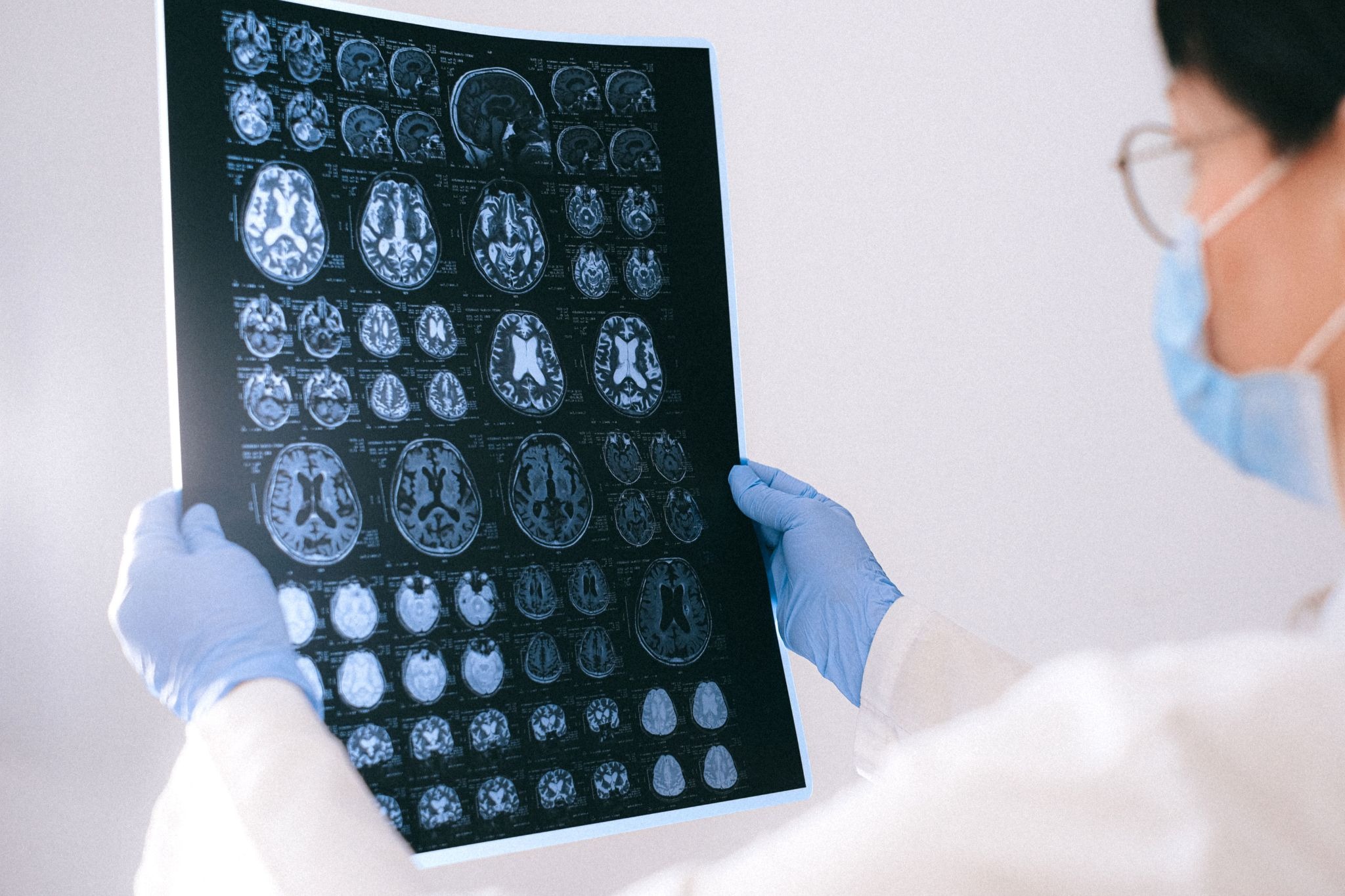
MRI scans contain information about the tumour’s appearance, such as its intensity, texture and the spatial arrangement of different features, which could provide further biological data about the tumour (e.g. water content). Researchers are investigating whether these imaging characteristics are associated with the tumour’s underlying molecular characteristics, including MGMT methylation.
Once the model has learned a reliable relationship between imaging patterns and MGMT status, it may be able to predict whether the tumour has MGMT promoter methylation on new, unseen MRI scans. MRI scans are already commonly used to assess patients with brain tumours. Hence, an AI model could potentially analyse scans obtained during routine clinical care without requiring an additional imaging procedure.
Researchers have also investigated whether incorporating other clinical patient data could improve the accuracy of these predictions. For example, patient characteristics, such as age and sex, alongside radiomic features–measurable characteristics extracted from medical images, such as aspects of tumour texture and appearance–could be fed into a predictive model as input [10, 12].
If the approach is successful, doctors may one day be able to upload the patient’s MRI scan to obtain information on the tumour’s MGMT methylation status. What currently requires an invasive procedure could potentially be performed computationally. This would, in turn, reduce the anxiety and stress that the patient experiences from having to face surgical risks, waiting for recovery, and experiencing anxiety from waiting [13].
Despite these advances, AI-based prediction of MGMT methylation remains an active area of research, and further validation is needed before these approaches can be incorporated into routine clinical care. Even if these tools eventually become suitable for clinical use, doctors may be hesitant to rely on these AI predictions when they cannot understand how those predictions were reached. A lot of these tools are described as “black boxes”–they can take information, such as an MRI scan, and produce a prediction, but it can be difficult to understand how the AI arrived at that prediction. This makes it harder for doctors to verify their reasoning [14]. A serious error could also have detrimental consequences, raising questions as to who should be held accountable [15].
This highlights the need for researchers to not only focus on making these solutions robust and reliable but also transparent and understandable to those who use them. This is just one example of how AI could transform modern medicine–a routine MRI scan may contain more information about a tumour than meets the eye, and researchers are now working to uncover it.
Article written by Nardiena Pratama, a PhD student from the AI for Biomedical Innovation Centre for Doctoral Training (CDT), University of Edinburgh.
Article edited by Priscilla Wong, a recent BSc Biological Sciences (Immunology) (Hons) graduate from the University of Edinburgh and Head Online News Editor for EUSci.
References:
- Wang, Y. et al. (2026). Lung cancer diagnosis from CT scans using artificial intelligence techniques: A global perspective. Clinics, 81, p.100930. doi:10.1016/j.clinsp.2026.100930.
- Fernandez-Mateos, J. et al. (2024). Tumor evolution metrics predict recurrence beyond 10 years in locally advanced prostate cancer. Nature Cancer, [online] 5(9), pp.1334–1351. doi:10.1038/s43018-024-00787-0.
- Louis, D.N. et al. (2016). The 2016 World Health Organization classification of tumors of the central nervous system: A summary. Acta Neuropathologica, [online] 131(6), pp.803–820. doi:10.1007/s00401-016-1545-1.
- Stupp, R. et al. (2005). Radiotherapy plus concomitant and adjuvant temozolomide for Glioblastoma. New England Journal of Medicine, [online] 352(10), pp.987–996. doi:10.1056/nejmoa043330.
- Taylor, O.G. et al. (2019). Glioblastoma multiforme: An overview of emerging therapeutic targets. Frontiers in Oncology, 9. doi:10.3389/fonc.2019.00963.
- Hegi, M.E. et al. (2005). MGMT gene silencing and benefit from temozolomide in glioblastoma. The New England Journal of Medicine, [online] 352(10), pp.997–1003. doi:10.1056/NEJMoa043331.
- Rosas-Alonso, R. et al. (2024). Evaluation of the clinical use of MGMT methylation in extracellular vesicle-based liquid biopsy as a tool for glioblastoma patient management. Scientific Reports, [online] 14(1). doi:10.1038/s41598-024-62061-8.
- Mansouri, A. et al. (2019). MGMT promoter methylation status testing to guide therapy for Glioblastoma: Refining the approach based on emerging evidence and current challenges. Neuro-oncology, [online] 21(2), pp.167–178. doi:10.1093/neuonc/noy132.
- Sai, V. et al. (2024). AI-driven estimation of O6 methylguanine-DNA-methyltransferase (MGMT) promoter methylation in Glioblastoma patients: A systematic review with bias analysis. Journal of Cancer Research and Clinical Oncology, 150(2). doi:10.1007/s00432-023-05566-5.
- Usuzaki, T. et al. (2024). Identifying key factors for predicting O6-Methylguanine-DNA methyltransferase status in adult patients with diffuse glioma: A multimodal analysis of demographics, radiomics, and MRI by variable vision transformer. Neuroradiology, 66(5), pp.761–773. doi:10.1007/s00234-024-03329-8.
- Restini, F.C.F. et al. (2024). AI tool for predicting MGMT methylation in glioblastoma for clinical decision support in resource limited settings. Scientific Reports, 14(1). doi:10.1038/s41598-024-78189-6.
- Ardakani, A.A. et al. (2022). Interpretation of radiomics features–A pictorial review. Computer Methods and Programs in Biomedicine, 215, p.106609. doi:10.1016/j.cmpb.2021.106609.
- Erbaş, D.H. et al. (2023). A qualitative study: Patients’ operating room experiences. Perioperative Care and Operating Room Management, 33, pp.100352–100352. doi:10.1016/j.pcorm.2023.100352.
- Rosenbacke, R. et al. (2024). How explainable artificial intelligence can increase or decrease clinicians’ trust in AI applications in health care: Systematic review. JMIR AI, [online] 3, pp.e53207-e53207. doi:10.2196/53207.
- Price II, W.N. et al. (2024). Liability for use of artificial intelligence in medicine. Research Handbook on Health, AI and the Law, [online] pp.150–166. doi:10.4337/9781802205657.ch09.
Leave a Reply